Arthrograms

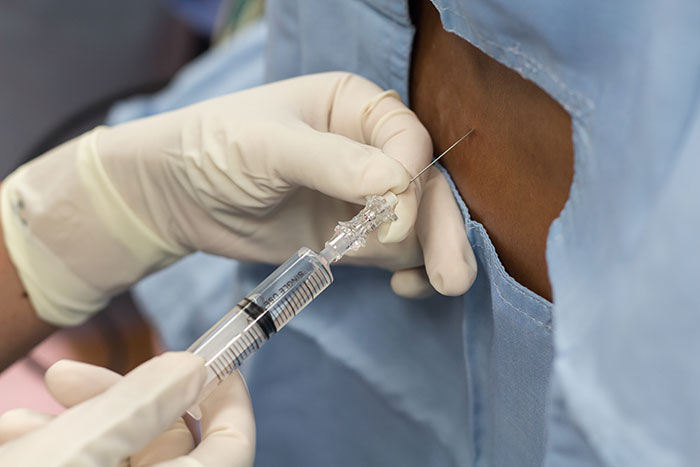
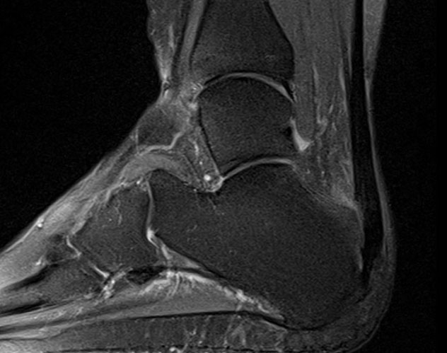
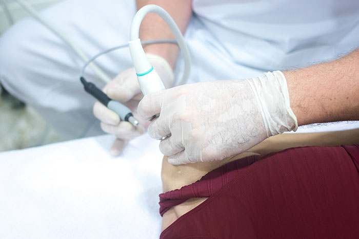
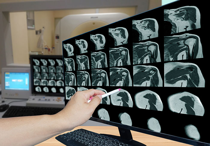
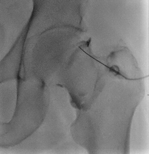
Using Image guidance (Fluroscopy or Ultrasound) contrast dye is injected into the joint. The patient is then subjected to a MRI or CT, which gives a better evaluation of the joint cartilage and other supporting structures, helping in the diagnosis.
The joints we commonly perform arthrograms on are Shoulders, Hips, wrists and elbows.
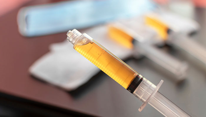
The same technique can you used for injection of Local Anasthesia, Steroid, hylouronic acid and PRP into the joint for treatment and diagnostic purpose.
Other procedures and diagnostics
Here at MSK, we perform a range of procedures with the help of imaging techniques, enhancing the efficiency, quality, and patient outcomes for a wide range of clients.
